To diagnose celiac disease in adults, we use the Catassi-Fasano criteria. These criteria state that at least four of the five diagnostic criteria for celiac disease must be met. Gluten should never be removed from the diet before the diagnostic workup is complete.
| At least 4 out of 5 | |
|---|---|
| 1 | Typical symptoms of celiac disease |
| 2 | Celiac disease specific IgA class antibodies with high titers |
| 3 |
Determination of HLA*-DQ2 and/or DQ8 *HLA: leukocyte histocompatibility antigen |
| 4 | Enteropathy compatible with CD in intestinal biopsy: includes Marsh-Oberhuber type 3 lesions, Marsh-Oberhuber type 1-2 associated with the presence of CD-specific antibodies with low/high titers, or Marsh-Oberhuber type 1-3 associated with subepithelial deposits of IgA and/or increased CD3+ TCRgd+ lymphocytes. |
| 5 | Response to DSG (histological response is required in patients with negative serology or associated with IgA deficiency). |
They are useful both for diagnosis and for monitoring and tracking the diet.
The antibodies we have available for diagnosis are:
- Tissue transglutaminase antibodies (tTG)
- These are the serological markers of choice due to their high sensitivity and specificity. When they are elevated five times above the upper limit of normal, the risk of false positives is very low. The higher the levels, the more specific they are for celiac disease.
- They are useful both for diagnosis and for monitoring and tracking the diet.
- Approximately 2.5% of patients with celiac disease present with a selective deficiency of total serum IgA. In cases where an IgA deficiency is confirmed, it is necessary to determine the presence of IgG class ATGT.
- Anti-endomysial antibodies
- They are less sensitive and more specific than ATGTs but expensive and not available at all centers. They are reserved for confirming some positive ATGT results.
Some people do not raise their antibody levels because they have an IgA deficiency, take immunosuppressants, or follow a low-gluten diet.
Furthermore, there is the concept of "seronegative celiac disease," which includes patients diagnosed with villous atrophy who respond to a gluten-free diet but have not shown elevated antibody levels in serology. It is important to note that only 30% of Marsh I and 85% of Marsh III patients are ATTG positive. Therefore, it is crucial to understand that a negative result for these markers does not definitively rule out the diagnosis.
CD is genetically determined by the configuration of the HLA (histocompatibility complex) system. The HLA-DQ2 (DQA1*05xx + DQB1*02xx) and/or HLA-DQ8 (DQA1*03 + DQB1*0302) genes encode the DQ2 and/or DQ8 heterodimers (antigen-presenting proteins) necessary for gluten to be presented to the immune system.
The presence of these genes (HLA-DQ2/DQ8) is necessary to develop the disease, but it is not sufficient, since these genetic factors are also present in high percentages of healthy individuals (30% of the total population). Therefore, celiac patients have these genotypes, but not all people with these genotypes will develop celiac disease.
- POSITIVE GENETIC STUDY = PREDISPOSITION TO DEVELOP CELIAC DISEASE
- A POSITIVE GENETIC TEST DOES NOT MEAN YOU HAVE CELIAC DISEASE
- NEGATIVE GENETIC STUDY = WE RULE OUT CELIAC DISEASE
Genetic testing is not necessary for all patients. It can be very useful in some situations:
- In patients with clinical suspicion but negative serology, a decision should be made regarding whether to perform duodenal biopsies. If there is no genetic risk, an alternative diagnosis should be considered; however, if a genetic risk is present, the need for a biopsy must be considered.
- To identify high-risk individuals among first-degree relatives and patients with autoimmune diseases. Individuals with a positive genetic predisposition should undergo regular clinical and analytical monitoring, as they may develop chronic malnutrition.
- The presence of several grades of enteropathy (from Marsh-Oberhuber 1 to 3) in a seronegative patient and a positive genetic test may indicate the presence of a seronegative AC.
- In patients who have removed gluten from their diet without a prior biopsy, who do not want to or cannot perform a gluten challenge test.
- In cases where there is no response to gluten withdrawal, to rule out a misdiagnosis.


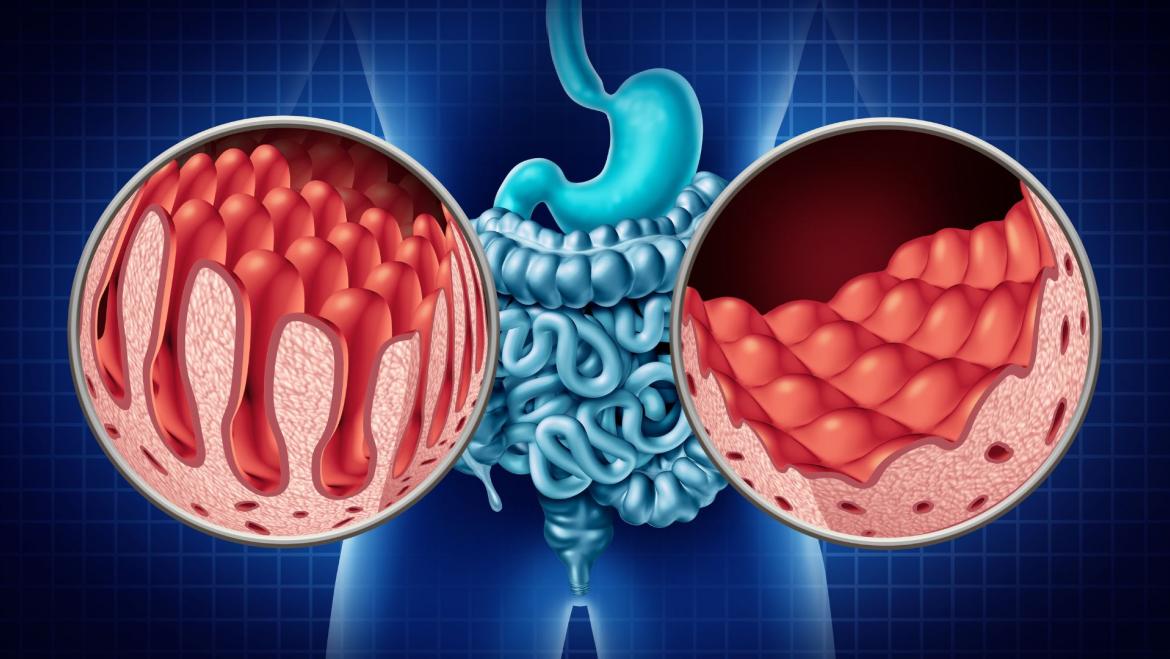
Histological examination is essential in the diagnostic strategy for celiac disease (especially in adults) and also allows for determining the severity of the lesion. Samples from the small intestine are obtained via upper gastrointestinal endoscopy under sedation while the patient is still consuming gluten.
An upper gastrointestinal endoscopy is an examination that allows for a detailed and complete view of the esophagus, stomach, and duodenum. It is performed by inserting a flexible tube through the mouth and inflating the stomach with air to improve visualization. During the procedure, a tissue sample (biopsy) can be taken for analysis, or any necessary treatments can be performed. It is a quick test, lasting approximately 10 minutes. The results of the endoscopy are available immediately afterward. If samples were taken, it will take about four weeks for a definitive diagnosis.
The test is usually performed with sedation, and therefore, despite being an invasive test, it is not uncomfortable and the patient is most likely not to notice the procedure and not to remember anything.
Preparation for upper digestive endoscopy
You must come FASTING. Important! If you are not fasting, we will NOT be able to perform the test. Your last meal, 8 hours before the test, must be LIGHT.
- 8 hours before the test: You cannot take anything except clear liquids (water, bowel preparation, herbal tea) or strong coffee and pulp-free juices (apple or white grape). Do not drink milk. Smoking is not recommended.
- 2 hours before the test: You must be fasting. Do NOT take anything (no water, no chewing gum, no bowel preparation, etc.).
If you have a pacemaker, ICD, drug allergies, or any heart or breathing problems, you should tell your doctor beforehand.
Endoscopy allows direct observation of macroscopic changes in the duodenal mucosa, including the fistoned pattern, reduction of folds, and nodularity. Although these changes are helpful in guiding biopsy sampling, they are not sensitive or specific enough for diagnosis, and biopsy is necessary. Because histological lesions may have an irregular distribution, multiple samples must be taken for intestinal biopsy (two from the duodenal bulb and four from the distal duodenum).


Important: Do not remove gluten from your diet before completing the diagnostic tests prescribed by the specialist team. Eliminating it prematurely may alter the results and lead to false negatives

The Marsh classification, modified by Oberhuber, establishes the histological spectrum of celiac disease. It is important to emphasize that celiac disease is NOT GRADES. All people with celiac disease are equally celiac and must follow the same strict diet. The fact that they have more symptoms or more intestinal damage does NOT mean they are less celiac. In all people with celiac disease, gluten causes intestinal damage, whether or not they develop symptoms.
| Segmented intraepithelial lymphocytes | Crypt hyperplasia | Villous atrophy | Types according to Marsh-Oberhuber |
|---|---|---|---|
| Yes | No | No | Tipus 1 |
| Yes | Yes | No | Tipus 2 |
| Yes | Yes | Yes (partial) | Tipus 3a |
| Yes | Yes | Yes (subtotal) | Tipus 3b |
| Yes | Yes | Yes (total) | Tipus 3c |
Samples from the small intestine for flow cytometry are also obtained from an upper digestive endoscopy under sedation.
Flow cytometry of duodenal biopsy allows obtaining an intraepithelial lymphogram. The complete celiac cytometric pattern (intraepithelial lymphocytes expressing T cell receptors – TCRs – γδ+ ≥ 10%, with a decrease in intraepithelial lymphocytes (CD3- ≤ 10%)) has a specificity close to 100% for the diagnosis of celiac disease, while the incomplete celiac cytometric pattern (intraepithelial lymphocytes with TCRs γδ+ ≥ 10%, but a normal count of intraepithelial lymphocytes (CD3- > 10%)) has a specificity of 91%. These lymphocytes have also been detected in mild enteropathy. This increase in TCRs γδ+ has not been observed in other common intestinal diseases, and therefore, the test may be useful for the differential diagnosis of gluten-sensitive enteropathy in individuals on a gluten-free diet.
This technique is very useful for the differential diagnosis of enteropathy with negative serology and in those classified as potential celiac patients (positive serology and absence of mucosal lesions or Marsh 1). The presence of a celiac lymphogram in the latter case necessitates a shared decision with the patient or their family regarding gluten withdrawal from the diet (assessing the symptomatic response) or, alternatively, close monitoring with periodic serological testing and repeating the biopsy at a later date.